
Abstract
Objective
Dysregulation of cell-cycle control mechanisms plays an important role in the development of many cancers, including cervical cancer. After the role of CDK 4/6 in cell cycle control and carcinogenesis was elucidated, CDK 4/6 inhibitors were developed. Ribociclib is a selective CDK4/6 inhibitor, and although its activity has been investigated in several malignancies, its potential role in cervical cancer remains incompletely defined. This study aimed to evaluate the chemosensitizing effects of ribociclib when combined with cisplatin and paclitaxel in a single human papillomavirus type 18 (HPV18)-positive cervical cancer cell line.
Material and Methods
For the experimental model, only the HPV18-positive human cervical adenocarcinoma-derived HeLa cell line (ATCC CCL-2) was used. Cells were treated for 24 hours with ribociclib (25 µM), cisplatin (100 µM) plus paclitaxel (2 µM), or cisplatin (100 µM) plus paclitaxel (2 µM) plus ribociclib (25 µM). Untreated cells served as controls. Cell viability was assessed using the 3-(4,5-dimethylthiazol-2-yl)-2,5-diphenyltetrazolium bromide (MTT) assay. Markers of angiogenesis, proliferation, apoptosis, and metastatic behavior were evaluated by immunocytochemical staining for VEGF, CDK4, CDK6, Ki-67, Bax, Bcl-2, caspase-3, E-cadherin, vimentin, fibronectin, and MMP-9.
Results
After 24 hours, cell viability was 48% in the cisplatin plus paclitaxel group and 55% in the cisplatin plus paclitaxel plus ribociclib group. Thus, the addition of ribociclib did not further reduce cell viability compared with chemotherapy alone. Addition of ribociclib to chemotherapy significantly reduced Bcl-2, vimentin, and MMP-9 expression and increased caspase-3 expression compared with chemotherapy alone. No significant additional reductions in VEGF or Ki-67, nor significant additional increases in Bax or E-cadherin, were observed with ribociclib.
Conclusion
In this single-cell-line, 24-hour in vitro model, ribociclib combined with cisplatin and paclitaxel was associated with changes in selected markers of apoptosis and metastatic behavior, despite not producing lower MTT-measured cell viability than chemotherapy alone. These findings constitute hypothesis-generating preclinical evidence and require validation in additional HPV18-positive and other cervical cancer models, in animal studies, and, ultimately, in clinical studies before any translational conclusions can be drawn.
Keywords: Cell cycle, cervical cancer, human papillomavirus, CDK4/6 inhibitor, ribociclib
INTRODUCTION
Cervical cancer remains a major global health problem. According to the most recent global cancer estimates, approximately 604,000 new cases and 280,000 deaths from cervical cancer were estimated worldwide in 2024; the disease remains a leading cause of cancer death among women globally. The burden is disproportionately concentrated in low- and middle-income countries, where approximately 90% of cases and deaths occur.
The development and progression of cervical carcinoma depend on various cellular genetic and epigenetic alterations, particularly abnormalities in cell-cycle checkpoints. Human papillomaviruses (HPVs) are the primary etiological agents of cervical carcinogenesis, with high-risk HPV types, particularly HPV16 and HPV18, being strongly associated with malignant transformation. HPVs promote carcinogenesis by interfering with normal cell-cycle control mechanisms. The oncogenic potential of HPV is largely mediated by E6 and E7 oncoproteins, which affect multiple targets involved in growth control. E6 contributes to malignant transformation through interactions with tumor suppressor p53 and pro-apoptotic BAK, whereas E7 inhibits the tumor suppressor retinoblastoma protein (Rb). These effects facilitate resistance to apoptosis, viral DNA replication, and continued cell-cycle progression.
Cyclin-dependent kinases (CDKs) are important regulators of cell-cycle progression and are among the pathways frequently dysregulated in cancer. Cell-cycle progression is controlled by CDKs (including CDK1, CDK2, CDK4, and CDK6), their activating cyclins, and CDK inhibitors. Among these mechanisms, the CDK4/6-cyclin D-Rb-E2F pathway is overactivated or dysregulated in many cancers. In this pathway, cyclin D forms complexes with CDK4 and CDK6, resulting in the phosphorylation of Rb. This releases E2F transcription factors and facilitates the transition from the G1 to the S phase of the cell cycle and subsequent cell proliferation. Alterations in CDKs, cyclins, and CDK inhibitors have also been implicated in cervical carcinogenesis and may contribute to uncontrolled proliferation and malignant transformation.
Selective CDK4/6 inhibitors have subsequently entered clinical practice. By inhibiting CDK4/6 kinase activity, these agents reduce Rb phosphorylation and suppress G1-to-S-phase progression., Ribociclib (R), palbociclib, and abemaciclib are approved CDK4/6 inhibitors used primarily in hormone receptor (HR)-positive, human epidermal growth factor receptor 2 (HER2)-negative breast cancer. Importantly, their clinical use is not restricted to postmenopausal patients or to use in combination with letrozole. For example, current U.S. Food and Drug Administration labeling for R includes adjuvant treatment of adults with HR-positive/HER2-negative stage II-III early breast cancer at high risk of recurrence, in combination with an aromatase inhibitor, as well as treatment of adults with HR-positive/HER2-negative advanced or metastatic breast cancer in combination with an aromatase inhibitor or fulvestrant. In pre/perimenopausal women and men receiving endocrine combinations, ovarian suppression with an luteinizing hormone-releasing hormone agonist is recommended according to clinical practice.-
Preclinical activity of CDK4/6 inhibitors has also been investigated in several solid tumors, including colorectal cancer, prostate cancer, sarcomas, glioblastoma, pancreatic ductal adenocarcinoma, melanoma, non-small-cell lung cancer, and esophageal adenocarcinoma. Importantly, previous work has already investigated R in cervical cancer models, including in vitro and in vivo systems. Therefore, the present study should be considered one of the early studies evaluating R-based strategies in cervical cancer, rather than the first study to investigate this question.
In cervical cancer, amplification of the cyclin D1 and CDK4 genes or overexpression of their proteins has been demonstrated; cyclin D1 overexpression has been reported as an independent adverse prognostic marker. These findings provide a biological rationale for investigating therapies targeting the CDK4/6-cyclin D-Rb-E2F pathway in cervical cancer. However, the response of HPV-positive cervical cancer models to CDK4/6 inhibition may be influenced by the underlying molecular context. Therefore, further preclinical evaluation is warranted.
The present study aimed to evaluate the effects of R combined with cisplatin and paclitaxel (CP + P) on a single HPV18-positive, cervical adenocarcinoma-derived HeLa cell line, with particular emphasis on angiogenesis, apoptosis, CDK4/6 expression, metastatic behavior, cell viability, wound closure, and cellular morphology.
MATERIAL AND METHODS
Materials
The HPV18-positive human cervical adenocarcinoma-derived HeLa cell line (ATCC CCL-2) was used as the sole experimental cell model. The cell line was commercially obtained from the Ministry of Agriculture and Forestry of the Republic of Türkiye. The following drugs were used: CP (10 mg/20 mL, Koçak Farma), R (HY-15777, MCE), and P (30 mg/5 mL, Koçak Farma). The drugs were supplied in liquid form and added to the cell culture medium.
Primary antibodies used for immunocytochemical staining included the following: E-cadherin (ab76055, Abcam), MMP-9 (sc-6841, Santa Cruz), vimentin (ab8978, Abcam), caspase-3 (sc-56053), Ki-67 (sc-23900), CDK4 (sc-166373, Santa Cruz), CDK6 (sc-7961, Santa Cruz), Bax (MA5-14003, Thermo Fisher Scientific), Bcl-2 (sc-7382, Santa Cruz), fibronectin (sc-8422, Santa Cruz), and VEGF (sc-7269, Santa Cruz). The study was approved by Afyonkarahisar University of Health and Sciences Clinical Research Ethics Committee (approval no: 2011-KAEK-2, date: 05.03.2021). Because no patient-derived data were used, patient informed consent was not applicable.
Cell Culture
HeLa cells were cultured in DMEM supplemented with 10% fetal bovine serum, 1% L-glutamine, and 1% penicillin-streptomycin, and were incubated at 37 °C in a humidified atmosphere containing 5% CO2 for 2 days. When cells reached approximately 80% confluence, they were allocated to four groups. Drug treatment was initiated at time 0, and cells were exposed to the treatment conditions for 24 hours ().
TABLE 1. Cell groups and administered treatments.
Group | Treatment |
Group 1 | Control (C): no drug treatment |
Group 2 | Ribociclib (R): ribociclib 25 µM |
Group 3 | Cisplatin + paclitaxel (CP + P): cisplatin 100 µM + paclitaxel 2 µM |
Group 4 | Cisplatin + paclitaxel + ribociclib (CP + P + R): cisplatin 100 µM + paclitaxel 2 µM + ribociclib 25 µM |
MTT Cell Toxicity and Viability Measurement
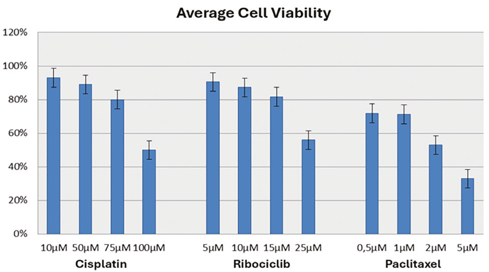
The MTT assay [3-(4,5-dimethylthiazol-2-yl)-2,5-diphenyltetrazolium bromide] was performed to evaluate the cytotoxic effects of different concentrations of CP, R, and P, and to determine the concentrations approximating the IC50. Concentration ranges were selected based on the literature and subsequently tested in HeLa cells. Cells were seeded in 96-well culture plates at 2 × 10⁴ cells/well. After cell adhesion, cells were treated for 24 hours with the following concentrations: CP, 5, 10, 50, 75, and 100 µM; R, 5, 10, 15, and 25 µM; and P, 0.5, 1, 2, and 5 µM.
At the end of the 24-hour treatment period, 20 µL/well of MTT solution (Sigma-Aldrich), prepared according to the manufacturer’s instructions, was added, and the plates were incubated at 37 °C for 4 hours. After removing the MTT solution, 200 µL of dimethyl sulfoxide was added to each well, and the plates were incubated on a shaker in the dark for 5 minutes. Absorbance was measured using a microplate reader (Shimadzu UV-1601) at 570-690 nm. Eight wells were prepared for each concentration, and the experiment was repeated three times. The mean results were used to select the treatment concentrations. The concentrations selected for subsequent experiments were CP (100 µM), R (25 µM), and P (2 µM) ().
Cytotoxic effective doses of CP, R and P in HeLa cells determined by MTT method.
The selected concentrations were subsequently applied to the CP + P and CP + P + R groups, and cell viability was assessed by MTT. This experiment was repeated seven times for each group.
CP + P + R: Cisplatin + paclitaxel + ribociclib; MTT: 3-(4,5-dimethylthiazol-2-yl)-2,5-diphenyltetrazolium bromide
Immunocytochemistry
Cells were expanded and seeded in 12-well culture plates. After adequate cell adhesion, CP, R, and P were administered to the experimental groups at concentrations determined by the MTT assay. After 24 hours, the culture medium was removed using a sterile pipette. Wells were washed once with phosphate-buffered saline (PBS) and fixed with 4% paraformaldehyde for 30 minutes. Permeabilization was performed with Triton X-100 on ice for 15 minutes. After washing, endogenous peroxidase activity was blocked with 3% H2O2. Wells were washed twice with PBS for 2 minutes each. After a 10-minute incubation in blocking solution, cells were incubated with the primary antibodies E-cadherin (1:250), MMP-9 (1:250), vimentin (1:250), caspase-3 (1:250), Ki-67 (1:250), CDK4 (1:250), CDK6 (1:250), Bax (1:250), Bcl-2 (1:250), fibronectin (1:250), and VEGF (1:250) for 1 hour at 37 °C. After washing with PBS, cells were treated with biotin for 30 minutes, followed by avidin for 30 minutes, as the secondary detection procedure. DAB was used as the chromogen. Nuclei were counterstained with Mayer’s hematoxylin and then mounted with a water-based mounting medium. Cells were evaluated using a light microscope at 20× objective magnification, and cell counts were performed using the NIS-Elements image analysis program (Japan).
Following immunocytochemical staining, H-scores were calculated as a semiquantitative measure for statistical analysis. For each group, 500 cells were counted per field. According to staining intensity, an H-score ranging from 0 to 500 was calculated using the formula described in the original study protocol: percentage of stained cells × staining intensity (0-5).- Immunocytochemical scoring was performed independently by two evaluators blinded to the treatment allocation. In cases of discrepancy, the final score was determined by consensus between the two evaluators.
Cell Migration and Wound Healing Assay
After HeLa cells reached 100% confluence, a line was drawn across the center of the monolayer using a sterile 10 µL pipette tip to create a wound area. Detached and dead cells were removed by changing the medium. Drug treatments were then applied to the experimental groups for 24 hours. Images were acquired using a phase-contrast microscope (ZEISS Axio Vert iLED) at 0, 6, 18, and 24 hours. Wound width and area were measured using the ZEN 3.3 Blue Edition (ZEN Lite) software, and time-dependent changes in wound area were compared statistically between groups.
Evaluation of Cell Morphology
Cell morphology was evaluated using phase-contrast microscopy (ZEISS Axio Vert iLED) immediately before treatment (0 hours) and 24 hours after treatment. Morphological changes were compared among the experimental groups.
Statistical Analysis
Statistical analyses were performed using SPSS version 28 (IBM Corp., Armonk, NY, USA). One-way analysis of variance was used for comparisons among groups, followed by Bonferroni correction for post-hoc comparisons. A two-sided p-value of <0.05 was considered statistically significant.
RESULTS
Effect of CP, P, and R on Cell Viability in HeLa Cells
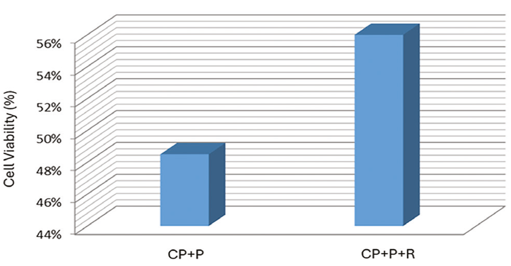
After 24 hours of treatment, MTT-measured cell viability was 48% in the CP + P group and 55% in the CP + P + R group. Thus, the addition of R did not result in lower cell viability measured by MTT than chemotherapy alone ().
Cytotoxic effects of CP + P and CP + P + R on HeLa cells determined by the MTT assay after 24 hours of treatment.
CP + P + R: Cisplatin + paclitaxel + ribociclib; MTT: 3-(4,5-dimethylthiazol-2-yl)-2,5-diphenyltetrazolium bromide
The slightly higher MTT-measured viability in the R-containing combination group should not be interpreted as evidence of increased cytotoxicity. One possible explanation is that some caspase-3-positive cells may have entered an apoptotic pathway without immediate loss of membrane integrity, and short-term MTT measurements may, therefore, not fully reflect the subsequent fate of these cells. Because the present experiment assessed viability only after 24 hours, longer treatment intervals would be required to determine whether R produces a delayed effect on cell viability.
Angiogenic Character of HeLa Cells after Treatment
VEGF expression was widespread and intense in the control group. After 24 hours of treatment, VEGF expression was significantly lower in the R, CP + P, and CP + P + R groups than in the control group (p<0.0001, p<0.0001, and p<0.0001, respectively) (). However, the addition of R to chemotherapy did not produce a further significant reduction in VEGF expression compared with chemotherapy alone.
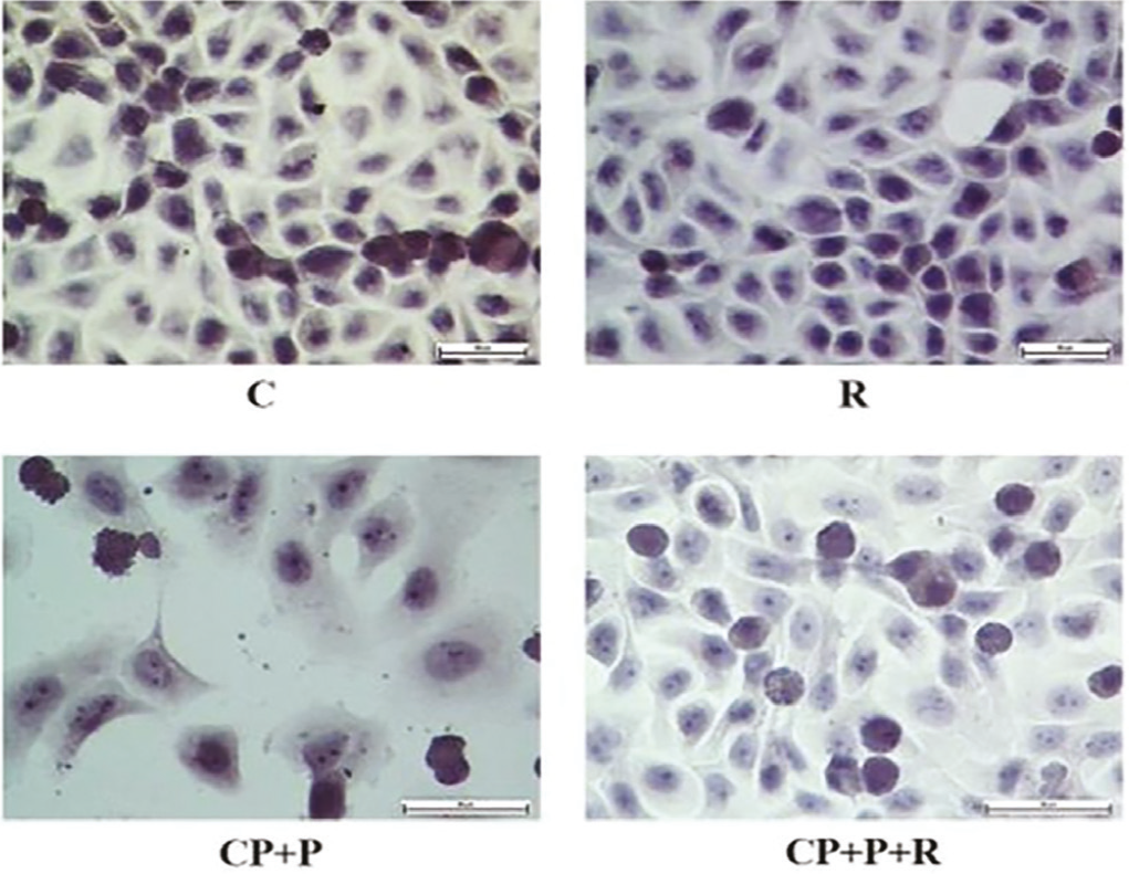
Immunocytochemical demonstration of VEGF protein expression in HeLa cells.
CP + P + R: Cisplatin + paclitaxel + ribociclib; VEGF: Vascular endothelial growth factor
CDK4 and CDK6 in HeLa Cells after Treatment
CDK4 and CDK6 staining was intense and widespread in both the control and CP + P groups, which exhibited similar staining patterns. The R and CP + P + R groups showed similar staining intensity and distribution, and both displayed significant reductions in staining intensity and in the number of positive cells compared with the control group (p<0.0001 for both comparisons) ().
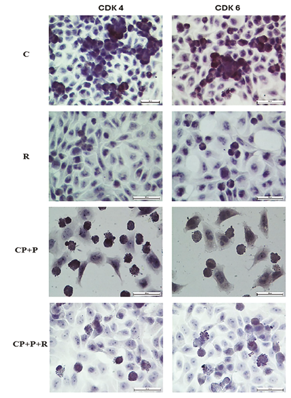
Immunocytochemical demonstration of CDK4 and CDK6 protein expression in HeLa cells.
Apoptosis and Cell Proliferation Markers in HeLa Cells after Treatment
Bax expression was similar between the control and R groups (p=1.0). Bax expression was increased in the CP + P and CP + P + R groups compared with the control group (p=0.001 and p=0.002, respectively). However, no significant difference was observed between CP + P and CP + P + R (p=1.0), indicating that adding R to chemotherapy did not further increase Bax expression.
Bcl-2 expression was highest in the control group. It was significantly reduced in the R and CP + P groups when compared with the control group (p<0.0001 for both), and the greatest reduction occurred in the CP + P + R group (p<0.0001). The addition of R to chemotherapy significantly reduced Bcl-2 expression compared with chemotherapy alone (p<0.0001).
Caspase-3 expression was increased in both the R and CP + P groups compared with the control group (p<0.0001 for both). Caspase-3 expression was highest in the CP + P + R group and was significantly higher than in the control group (p<0.0001). The addition of R to chemotherapy significantly increased caspase-3 expression compared with chemotherapy alone (p=0.002).
Ki-67 expression was intense and widespread in the control group but significantly reduced in the R, CP + P, and CP + P + R groups (p<0.0001 for all). However, the addition of R to chemotherapy did not produce a significant additional reduction in Ki-67 expression compared with chemotherapy alone (p=1.0) ().
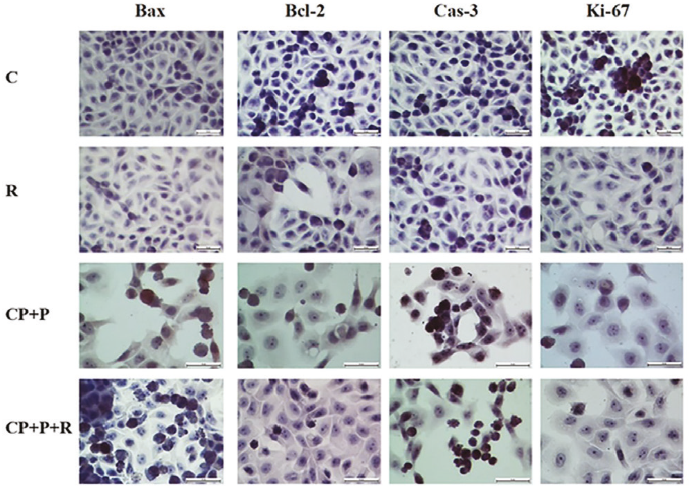
Immunocytochemical demonstration of apoptotic markers (Bax, Bcl-2, and caspase-3) and the proliferation marker Ki-67 in HeLa cells.
CP + P + R: Cisplatin + paclitaxel + ribociclib
Evaluation of Metastatic Behavior of Cells
Fibronectin expression increased modestly in the treatment groups compared with the control group. No significant difference was observed between the control and R groups (p=1.0); fibronectin expression was significantly higher in the CP + P and CP + P + R groups than in the control group (p=0.001 and p<0.0001, respectively). No significant difference was observed between CP + P and CP + P + R (p=0.304).
Vimentin expression was widespread and intense in the control group, and was significantly reduced in the R, CP + P, and CP + P + R groups compared with control (p<0.0001 for all). Furthermore, vimentin expression was significantly lower in the CP + P + R group than in the CP + P group (p<0.0001).
E-cadherin expression was increased in all treatment groups compared with the control group, with the greatest increase observed in the CP + P + R group (p<0.0001 for all comparisons with the control). However, the difference between CP + P and CP + P + R was not statistically significant (p=0.097).
MMP-9 expression was highest in the control group and lower in the R, CP + P, and CP + P + R groups (p<0.0001 for all comparisons with control). The greatest reduction occurred in the CP + P + R group, and MMP-9 expression was significantly lower in the CP + P + R group than in the CP + P group (p<0.0001) ().
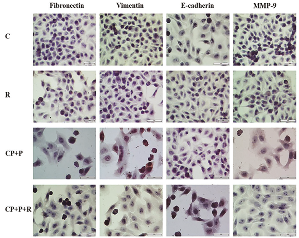
Immunocytochemical demonstration of fibronectin, vimentin, E-cadherin, and MMP-9 expression in HeLa cells.
CP + P + R: Cisplatin + paclitaxel + ribociclib
A wound-healing assay was performed to assess treatment-related changes in cell migration. Images were obtained at 0, 6, 18, and 24 hours, and wound areas were compared among groups (). Wound areas were similar among groups at baseline. At 24 hours, the wound was completely closed in the control group, whereas closure in the CP + P + R group was significantly lower (p<0.001). At the 24th hour, wound closure in the CP + P group was significantly less than in the CP + P + R group (p=0.011). These findings indicate that R did not further delay wound closure beyond that observed with the chemotherapy combination.
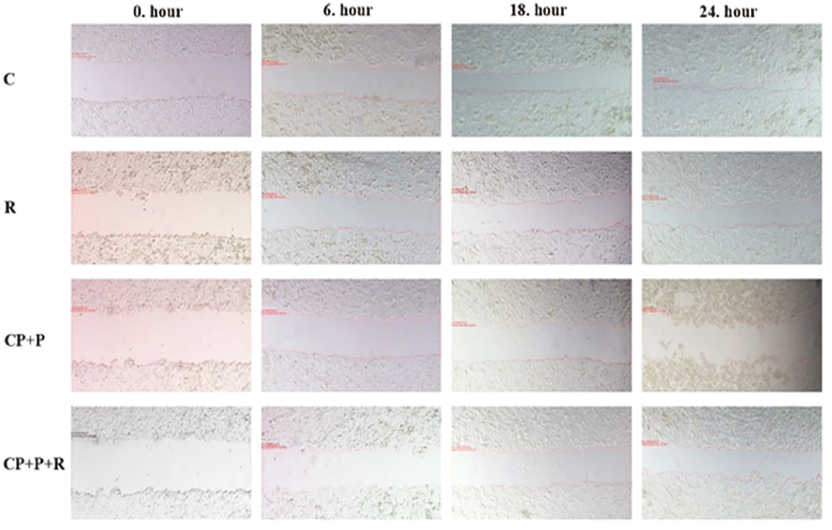
Wound-healing assay images of HeLa cells obtained at the indicated time points after treatment. Scale bar, 200 µm.
CP + P + R: Cisplatin + paclitaxel + ribociclib
Morphological Changes in Cells
Cells were visualized by phase-contrast microscopy after 24 hours of treatment (). Compared with the untreated control group, the CP + P and CP + P + R groups showed substantial morphological changes. Cells in the control and R groups were generally similar in cell size, nuclear and cytoplasmic appearance, and overall shape. In contrast, cells treated with CP + P or CP + P + R showed reduced adherence, a smaller, more rounded appearance, disrupted nuclear and cytoplasmic morphology, prominent vacuolization, and nuclear fragmentation.
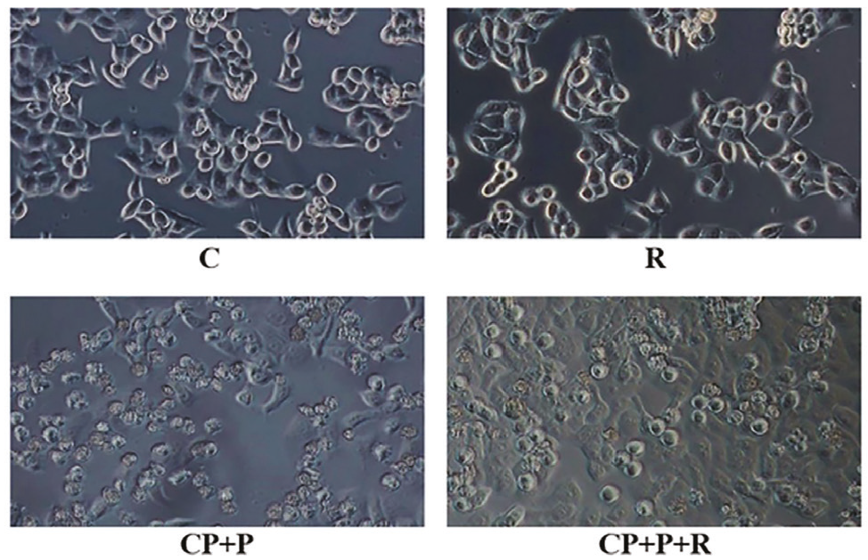
Morphological changes in HeLa cells after 24 hours of treatment. Scale bar, 200 µm.
CP + P + R: Cisplatin + paclitaxel + ribociclib
DISCUSSION
Apoptosis is an important mechanism for clearing away damaged or abnormal cells, whereas escape of apoptosis contributes to cancer-cell survival and tumor progression. Anti-apoptotic Bcl-2 and pro-apoptotic Bax are key regulators of apoptotic cell death. Following a death signal, Bax promotes mitochondrial outer-membrane permeabilization and cytochrome c release, which contribute to apoptosome formation and caspase activation. Caspase-3 has a vital role in this cascade and contributes to the morphological and biochemical changes of apoptosis.,
In HPV-positive human cervical cancer cell line, P has been reported to activate the mitochondria-derived apoptotic pathway by down-regulating Bcl-2 through cytochrome c release. Overexpression of Bcl-2 can contribute to resistance to chemotherapy and radiotherapy by limiting mitochondrial apoptotic signaling. In the present study, the addition of R to CP + P significantly reduced Bcl-2 levels and increased caspase-3 levels compared with chemotherapy alone. These findings are consistent with increased activation of apoptotic signaling and may suggest that adding R to chemotherapy could improve treatment outcomes; however, because cell viability was not reduced in the combination group in the MTT assay, and because the study was limited to a single cell line with 24 hours of drug exposure, these results should be interpreted as hypothesis-forming observations rather than evidence of improved treatment efficacy.
Modification of the extracellular matrix by neoplastic cells, including matrix degradation and altered synthesis of extracellular-matrix components, plays an important role in tumor dissemination. MMPs are the most important proteases involved in the degradation of the extracellular matrix and play a key role in invasion and metastasis. Expression of MMPs enables the release of growth factors bound to the extracellular matrix and/or to membranes, thereby providing a microenvironment conducive to extracellular matrix remodeling and primary tumor formation. MMP-9 expression has been reported to be increased in cervical cancer and associated with clinicopathological characteristics and prognosis.,
Epithelial-mesenchymal transition (EMT), initially described during embryonic development, also contributes to tumor-cell migration, invasion, and metastasis.,Loss of E-cadherin and increased expression of mesenchymal markers such as N-cadherin, vimentin, and fibronectin are commonly used as indicators of EMT. E-cadherin is an intercellular adhesion molecule, and loss of E-cadherin is associated with increased invasion and tumor stage. Vimentin is a protein of intermediate filaments, and its increased expression promotes migration, invasion and metastasis of tumors, while it also contributes to the development of infiltration and metastasis by reducing E-cadherin-mediated intercellular adhesion.,In cervical squamous cell carcinoma, associations between decreased E-cadherin, increased vimentin, and overall survival have been reported.
Previous clinical and preclinical studies have explored CDK4/6 inhibition in both HPV-positive and HPV-negative malignancies. In a phase 1 study of palbociclib plus cetuximab in recurrent head and neck squamous cell carcinoma, prolonged time to progression was observed in two HPV-positive patients, although the mechanism was uncertain. In a phase 2 study of palbociclib plus cetuximab in cetuximab-resistant HPV-positive oropharyngeal squamous cell carcinoma, the primary endpoint was not met. Similarly, a study of palbociclib plus cetuximab in platinum-resistant HPV-negative head and neck cancer did not meet its primary endpoint. While the underlying mechanism for the failure to meet the primary endpoint in these two studies is unclear, one reason could be that patients did not receive chemotherapy, and another could be the sensitivity of the treated tumor to the CDK 4/6 inhibitor. In our study, the significant reduction in vimentin and MMP-9 observed when R was added to chemotherapy raises the hypothesis that R may influence chemotherapy sensitivity in HPV-18-positive cervical cancer cells via an off-target mechanism. These findings illustrate the uncertainty surrounding CDK4/6 inhibition in HPV-associated cancers and the importance of tumor context and treatment combinations.
Our findings showed that adding R to CP + P significantly reduced vimentin and MMP-9 expression, increased caspase-3, and further reduced Bcl-2. These changes may indicate altered apoptotic and metastatic behavior pathways. However, the present data do not establish increased chemosensitivity, delayed chemotherapy resistance, or clinical efficacy. In particular, the absence of lower MTT-measured viability in the combination group compared with chemotherapy alone argues against making a direct claim of enhanced cytotoxicity based on the current experiment. The observed marker changes, therefore, provide a rationale for further investigation rather than sufficient evidence to support a treatment recommendation.
Study Limitations
Several limitations should be considered. First, only one cell line was studied. HeLa is an HPV18-positive cervical adenocarcinoma-derived cell line; therefore, the findings cannot be generalized to all HPV-positive cervical cancers, other HPV genotypes, or other cervical histological subtypes. Second, the concentrations used in the present experiments (CP 100 µM, P 2 µM, and R 25 µM) are relatively high and may not directly correspond to clinically achievable tumor exposures. Third, the treatment duration was limited to 24 hours; longer exposure periods (e.g., 48 or 72 hours) were not evaluated. Fourth, molecular markers of angiogenesis, apoptosis, proliferation, and metastatic behavior were assessed using a semiquantitative immunocytochemical approach; the findings were not independently validated by quantitative RT-PCR or Western blotting. Finally, R, alone and in combination with chemotherapy, was not evaluated in an in vivo cervical cancer model. Additional experiments using multiple HPV18-positive and other HPV-positive and -negative cervical cancer cell lines, employing longer treatment schedules, applying quantitative molecular assays, and involving animal models are needed.
The current results should therefore be viewed as preliminary in vitro evidence supporting further investigation of R in combination with standard cytotoxic agents in selected cervical cancer models. Confirmation in animal models and subsequently in well-designed clinical studies will be necessary before any translational or therapeutic conclusions can be drawn.
CONCLUSION
This study is among the early preclinical investigations of R combined with chemotherapy in an HPV18-positive cervical adenocarcinoma-derived cell line. After 24 hours of treatment, the addition of R to CP + P was associated with significantly lower expression of Bcl-2, vimentin, and MMP-9, and higher expression of caspase-3 compared with chemotherapy alone, but it did not reduce MTT-measured cell viability below that observed with chemotherapy alone. These findings provide hypothesis-generating evidence for changes in apoptotic markers and markers of metastatic behavior following exposure to R. Further validation across additional cervical cancer cell lines, extended treatment schedules, quantitative molecular assays, animal models, and clinical studies is required before the combination can be considered a potential therapeutic strategy.
Ethics
Ethics Committee Approval: The study was approved by Afyonkarahisar University of Health and Sciences Clinical Research Ethics Committee (approval no: 2011-KAEK-2, date: 05.03.2021).
Informed Consent: Because no patient-derived data were used, patient informed consent was not applicable.
Footnotes
Authorship Contributions
Surgical and Medical Practices: S.E.D., M.B., H.D., Concept: S.E.D., M.B., Design: S.E.D., M.B., Data Collection or Processing: S.E.D., M.B., F.F., H.D., Analysis or Interpretation: S.E.D., M.B., F.F., H.D., Literature Search: S.E.D., M.B., F.F., H.D., Writing: S.E.D., M.B., F.F., H.D.
Conflict of Interest
No conflict of interest was declared by the authors.
Financial Disclosure
This study was supported by the Turkish Society of Medical Oncology under grant number P-TTOD-2021-146.
References
Sung H, Filho AM, Laversanne M, et al. Global cancer statistics 2024: GLOBOCAN estimates of incidence and mortality worldwide for 34 cancers in 186 countries. CA Cancer J Clin. 2026;76(4):e70090.
Kim YT, Zhao M. Aberrant cell cycle regulation in cervical carcinoma. Yonsei Med J. 2005;46(5):597-613.
Milde-Langosch K, Riethdorf S. Role of cell-cycle regulatory proteins in gynecological cancer. J Cell Physiol. 2003;196(2):224-244.
Hu Z, Ma D. The precision prevention and therapy of HPV-related cervical cancer: new concepts and clinical implications. Cancer Med. 2018;7(10):5217-5236.
Hanahan D, Weinberg RA. Hallmarks of cancer: the next generation. Cell. 2011;144(5):646-674.
Xiong Y, Li T, Assani G, et al. Ribociclib, a selective cyclin D kinase 4/6 inhibitor, inhibits proliferation and induces apoptosis of human cervical cancer in vitro and in vivo. Biomed Pharmacother. 2019;112:108602.
Musgrove EA, Caldon CE, Barraclough J, Stone A, Sutherland RL. Cyclin D as a therapeutic target in cancer. Nat Rev Cancer. 2011;11(8):558-572.
Hamilton E, Infante JR. Targeting CDK4/6 in patients with cancer. Cancer Treat Rev. 2016;45:129-138.
Beaver JA, Amiri-Kordestani L, Charlab R, et al. FDA approval: palbociclib for the treatment of postmenopausal patients with estrogen receptor-positive, HER2-negative metastatic breast cancer. Clin Cancer Res. 2015;21(21):4760-4766.
Finn RS, Crown JP, Lang I, et al. The cyclin-dependent kinase 4/6 inhibitor palbociclib in combination with letrozole versus letrozole alone as first-line treatment of oestrogen receptor-positive, HER2-negative, advanced breast cancer (PALOMA-1/TRIO-18): a randomised phase 2 study. Lancet Oncol. 2015;16(1):25-35.
Kwapisz D. Cyclin-dependent kinase 4/6 inhibitors in breast cancer: palbociclib, ribociclib, and abemaciclib. Breast Cancer Res Treat. 2017;166(1):41-54.
FDA. KISQALI (ribociclib) tablets: prescribing information [Internet]. Silver Spring (MD): U.S. Food and Drug Administration; 2025.
Schettini F, De Santo I, Rea CG, et al. CDK 4/6 inhibitors as single agent in advanced solid tumors. Front Oncol. 2018;8:608.
Cheung TH, Yu MM, Lo KW, Yim SF, Chung TK, Wong YF. Alteration of cyclin D1 and CDK4 gene in carcinoma of uterine cervix. Cancer Lett. 2001;166(2):199-206.
Keller E, Goldman RD. Light microscopy. In: Spector DL, Goldman RD, editors. Basic methods in microscopy: protocols and concepts from cells: a laboratory manual. China: Cold Spring Harbor Laboratory Press; 2006. p. 1-42.
Nicholson RI, Bouzubar N, Walker KJ, et al. Hormone sensitivity in breast cancer: influence of heterogeneity of oestrogen receptor expression and cell proliferation. Eur J Cancer. 1991;27(7):908-913.
McCarty KS Jr, Miller LS, Cox EB, Konrath J, McCarty KS Sr. Estrogen receptor analyses. Correlation of biochemical and immunohistochemical methods using monoclonal antireceptor antibodies. Arch Pathol Lab Med. 1985;109(8):716-721.
McCarty KS Jr, Szabo E, Flowers JL, Cox EB, Leight GS, Miller L, et al. Use of a monoclonal anti-estrogen receptor antibody in the immunohistochemical evaluation of human tumors. Cancer Res. 1986;46(8 Suppl):4244s-4248s.
Tadrous PJ. Breast. In: Diagnostic criteria handbook in histopathology: a surgical pathology vade mecum. England: John Wiley & Sons; 2007. p. 258-266.
Joseph L, Srinivasan KK. Triacontanoic ester of 5’’-hydroxyjustisolin: Tumour suppressive role in cervical cancer via Bcl-2, BAX and caspase-3 mediated signalling. Toxicol Rep. 2019;6:1198-1205.
Tjalma WA, Weyler JJ, Bogers JJ, et al. The importance of biological factors (bcl-2, bax, p53, PCNA, MI, HPV and angiogenesis) in invasive cervical cancer. Eur J Obstet Gynecol Reprod Biol. 2001;97(2):223-230.
Dang YP, Yuan XY, Tian R, Li DG, Liu W. Curcumin improves the paclitaxel-induced apoptosis of HPV-positive human cervical cancer cells via the NF-κB-p53-caspase-3 pathway. Exp Ther Med. 2015;9(4):1470-1476. Retraction in: Exp Ther Med. 2025;30(2):152.
Yim EK, Tong SY, Ho EM, Bae JH, Um SJ, Park JS. Anticancer effects on TACC3 by treatment of paclitaxel in HPV-18 positive cervical carcinoma cells. Oncol Rep. 2009;21(2):549-557.
Goldberg I, Davidson B, Lerner-Geva L, et al. Expression of extracellular matrix proteins in cervical squamous cell carcinoma--a clinicopathological study. J Clin Pathol. 1998;51(10):781-785.
Li XW, Tuergan M, Abulizi G. Expression of MAPK1 in cervical cancer and effect of MAPK1 gene silencing on epithelial-mesenchymal transition, invasion and metastasis. Asian Pac J Trop Med. 2015;8(11):937-943.
Zhu D, Ye M, Zhang W. E6/E7 oncoproteins of high risk HPV-16 upregulate MT1-MMP, MMP-2 and MMP-9 and promote the migration of cervical cancer cells. Int J Clin Exp Pathol. 2015;8(5):4981-4989.
Yang J, Nakamura I, Roberts LR. The tumor microenvironment in hepatocellular carcinoma: current status and therapeutic targets. Semin Cancer Biol. 2010;21:35-43.
Ogunwobi OO, Liu C. Therapeutic and prognostic importance of epithelial-mesenchymal transition in liver cancers: insights from experimental models. Crit Rev Oncol Hematol. 2012;83 319-328.
Wang H, Zhou Q. [E-cadherin/beta-catenin and the invasion and metastasis of lung cancer]. Zhongguo Fei Ai Za Zhi. 2010;13(3):254-259. Chinese.
Yu JQ, Zhou Q, Zheng YF, Bao Y. Expression of vimentin and Ki-67 proteins in cervical squamous cell carcinoma and their relationships with clinicopathological features. Asian Pac J Cancer Prev. 2015;16(10):4271-4275.
Cheng Y, Zhou Y, Jiang W, et al. Significance of E-cadherin, β-catenin, and vimentin expression as postoperative prognosis indicators in cervical squamous cell carcinoma. Hum Pathol. 2012;43(8):1213-1220.
Michel L, Ley J, Wildes TM, et al. Phase I trial of palbociclib, a selective cyclin dependent kinase 4/6 inhibitor, in combination with cetuximab in patients with recurrent/metastatic head and neck squamous cell carcinoma. Oral Oncol. 2016;58:41-48.
Oppelt P, Ley JC, Worden F, et al. Palbociclib and cetuximab in cetuximab-resistant human papillomavirus-related oropharynx squamous-cell carcinoma: a multicenter phase 2 trial. Oral Oncol. 2021;114:105164.
Adkins DR, Lin JC, Sacco A, et al. Palbociclib and cetuximab compared with placebo and cetuximab in platinum-resistant, cetuximab-naïve, human papillomavirus-unrelated recurrent or metastatic head and neck squamous cell carcinoma: a double-blind, randomized, phase 2 trial. Oral Oncol. 2021;115:105192.