
Abstract
Immune checkpoint inhibitors (ICIs) have transformed the field of cancer immunotherapy and led to substantial improvements in patient outcomes across various malignancies. Neurological toxicities arising from ICI treatment represent a heterogeneous group of complications that manifest across a broad spectrum, ranging from mild symptoms to life-threatening conditions. The present article reviews patients receiving ICI treatment and identifies neurological adverse events observed across all ICI therapeutic modalities. Data were retrospectively evaluated from 500 cancer patients who received immunotherapy treatment between 2020-2022 at Koç University Hospital Medical Oncology Outpatient Clinic. Eight patients (1.6%) who developed immunotherapy-related neurologic side effects were included in the analysis. Demographic and clinicopathologic characteristics, along with laboratory results, were extracted from the medical oncology outpatient clinic database. In this study, 89% (7/8) of the patients were male, with a median age of 59 years (range 44-79). The most common cancer types observed were small cell lung cancer (n=2) and renal cell carcinoma (n=2). A case study is also presented of a patient who developed neurotoxicity following immunotherapy. Immunotherapy emergence has marked substantial advancement in cancer treatment approaches, although neurological side effects require close monitoring. Recognition of diverse neurological complications associated with ICIs and their potential severity remains essential for clinical practice.
Keywords: Immune related adverse events; immune checkpoint inhibitors; immunotherapy; neurological adverse events; neurological toxicities
INTRODUCTION
Immune checkpoint inhibitors (ICIs) have transformed the field of cancer immunotherapy and led to substantial improvements in patient outcomes across various malignancies. These inhibitors, targeting programmed cell death protein 1 (PD-1), programmed death-ligand 1 (PD-L1), and cytotoxic T-lymphocyte-associated protein 4 (CTLA-4), demonstrate efficacy in treating multiple cancers, including melanoma, non-small cell lung cancer, and renal cell carcinoma.- However, as the clinical use of ICIs has expanded, an increasing number of immune-related adverse events (irAEs) have been reported, including a variety of neurological complications.-
Neurological toxicities arising from ICI treatment represent a heterogeneous group of complications that manifest across a broad spectrum, ranging from mild symptoms to life-threatening conditions., , Such toxicities affect both central and peripheral nervous systems, manifesting as encephalitis, myasthenia gravis (MG), Guillain-Barré syndrome (GBS), and additional neurological syndromes., , The median onset time for immunotherapy-related neurotoxicity is established at four weeks, with occurrence possible between one week to 68 weeks. While neurological irAEs exhibit lower incidence compared to other irAEs, these events potentially result in significant morbidity and mortality., ,
The precise mechanisms underlying neurological irAE development remain incompletely understood, though attribution to immune system dysregulation caused by checkpoint inhibition has been proposed. Considering the expanding utilization of ICIs in cancer treatment, understanding clinical manifestations, diagnostic approaches, and management strategies for neurological complications becomes essential for clinicians and researchers., ,
The present article reviews patients receiving ICI treatment who developed neurological adverse events. Clinical spectrum, treatment approaches, and outcomes of ICI-related neurotoxicity are presented through a case-based methodology.
METHODS
Data were retrospectively evaluated from 500 cancer patients who received immunotherapy treatment between 2020-2022 at Koç University Hospital Medical Oncology Outpatient Clinic. Eight patients (1.6%) who developed immunotherapy-related neurologic side effects were included in the analysis. Demographic characteristics, clinicopathologic features, and laboratory results were extracted from the medical oncology outpatient clinic database.
Consent to Participate
Patient data were obtained retrospectively from medical records following acquisition of written informed consent from patients or designated relatives.
RESULTS
The study population comprised 89% (7/8) male patients, with a median age of 59 years (range: 44-79). Small cell lung cancer (n=2) and renal cell carcinoma (n=2) represented the most frequent cancer types. Cranial radiotherapy was administered to two patients with brain metastases. Atezolizumab treatment was received by four patients (50%). The median time to side effect onset was documented at 10.5 weeks (range: 1-95 weeks). Disease progression or infection resulted in mortality for five patients (63%) during the follow-up period. Clinical data are presented in .
TABLE 1. Demographic and clinical findings, treatment characteristics, and outcomes.
Patients | Diagnosis | Treatment | Immunotherapy | Toxicity | Treatment | Status |
|---|---|---|---|---|---|---|
44 years old female | Breast cancer | IO+ChT | Atezolizumab | Transverse myelitis | Oral methylprednisolone 1 mg/kg/day for 5 days and then oral 60 mg/day maintance - the steroid dose was tapered by 8 mg every five days. During the follow-up period, the dose was reduced to 16 mg/day and continued as maintenance therapy | Symptoms did not improve. The patient passed away due to infection |
44 years old male | SCLC | IO+ChT | Atezolizumab | Demyelinating disease | IV methylprednisolone 1000 mg/day for 7 days and then oral steroid 60 mg/day maintance. The steroid dose was tapered by 8 mg every five days. During the follow-up period, the dose was reduced to 8 mg/day and continued as maintenance therapy | Slight improvement in symptoms. The patient passed away due to disease progression |
68 years old male | Renal cell carcinoma | IO | Nivolumab | Encephalopathy | Oral methylprednisolone 1 mg/kg/day for 5 days and then oral 60 mg/day maintance- Steroid treatment was tapered by 12 mg/day every 5 days and discontinued within a month | Slight improvement in symptoms. The patient passed away due to aspiration pneumonia |
61 years old male | Malignant melanoma | IO+IO | Nivolumab plus Ipilimumab | Encephalopathy | IV methylprednisolone 1 mg/kg/day for 5 days and then oral 60 mg/day maintance +IVIG 33gr, D1-4. Steroid treatment was tapered by 12 mg/day every 5 days and discontinued within a month | Symptoms improved, and steroid dosage was tapered off and eventually discontinued. The patient continued with single-agent IO treatment. |
57 years old male | SCLC | IO+ChT | Atezolizumab | Encephalopathy | Oral methylprednisolone was administered at 16 mg/day for five days, after which the dose was reduced by 4 mg every three days, with the treatment discontinued within three weeks | Symptoms improved, and the steroid dosage was tapered off and eventually discontinued. Not avaliable follow-up data |
58 years old male | Larynx tumor | IO+Cht | Pembrolizumab | Acute disseminated polineuroradiculopaty with miyasthenic syndrome | Oral steroid 30 mg/day + IVIG 27 gr, D 1-5 and then iv pulse metyl prednisone 250 mg/day/five days, pyridostigmine 4x60 mg. The prednisolone dose was reduced to 30 mg and and continued as maintenance therapy | Symptoms improved, and the steroid dosage was tapered off but not discontinued. The patient passed away due to disease progression |
79 years old male | Renal cell carcinoma | IO | Nivolumab | Encephalopathy | Oral methylprednisolone was administered at 16 mg/day for five days, after which the dose was reduced by 4 mg every three days, with the treatment discontinued within three weeks | Alive -Not avaliable follow-up data |
68 years old male | Lung adenocarcinoma | IO+ChT | Atezolizumab | Demyelinating disease | Intravenous (IV) methylprednisolone was administered at 1000 mg/day for 5 days, followed by oral steroids at a maintenance dose of 60 mg/day. IVIG, at a total dose of 27 g, was administered over days 1-4. The steroid dose was tapered by 8 mg every five days. During the follow-up period, the dose was reduced to 16 mg/day and continued as maintenance therapy | Symptoms did not improve. The patient passed away due to infection |
SCLC: Small cell carcinoma; ChT: Chemotherapy; IO: Immunotherapy; IVIG: Intravenous immunglobuline.
CASE REPORT
A 59-year-old male patient was diagnosed with laryngeal cancer in February 2021. After completing 28 days of radiotherapy, a salvage laryngectomy was performed in January 2022 due to a local recurrence. Postoperative pathology indicated T4N0M0, and follow-up assessments were scheduled every three months. In July 2022, chemotherapy was initiated for a recurrent lesion that was considered unsuitable for surgical resection. Given a 60% PD-L1 expression level, treatment with a combination of cisplatin, 5-FU, and pembrolizumab was planned.
One month after completing two treatment cycles, the patient presented to the emergency department with right leg weakness, left arm weakness, and ptosis in the right eye. Neurological assessment revealed multiple cranial nerve paralysis manifesting as decreased eye squeezing, rightward tongue deviation and reduced tongue movements. Motor examination demonstrated extremity weakness (proximal more prominent than distal) with bilaterally absent deep tendon reflexes and plantar skin responses. Cranial and cervical magnetic resonance imaging (MRI) with electromyography examination suggested acute disseminated polyneuroradiculopathy ().
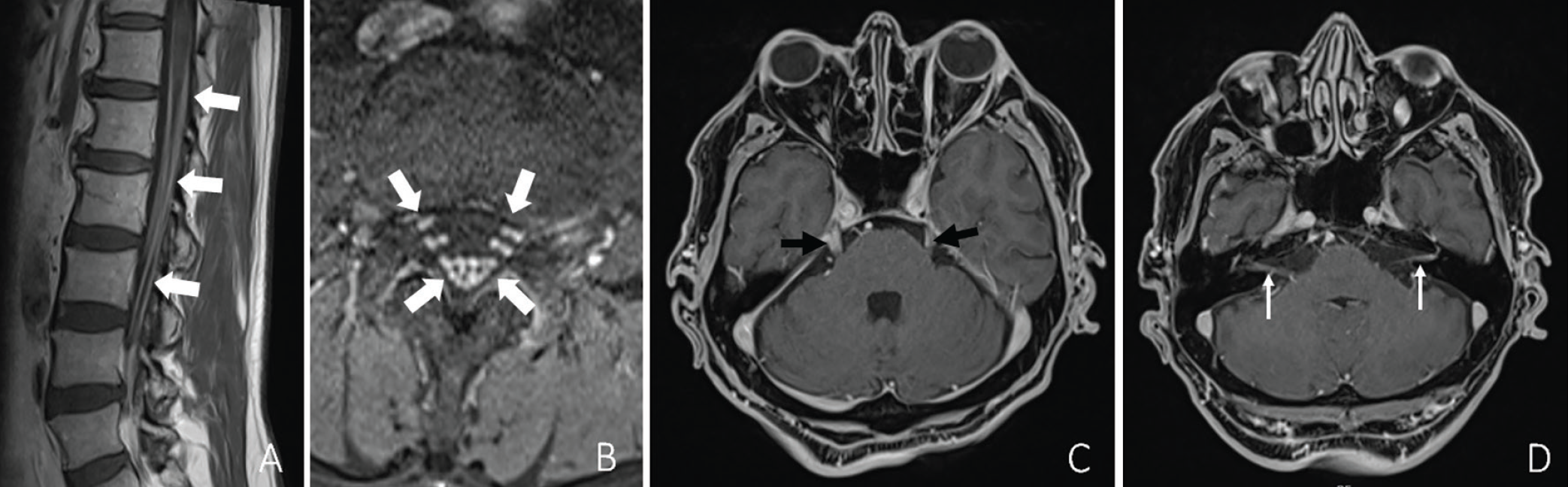
Admission to neurology service followed with preliminary diagnosis of acute disseminated polyneuroradiculopathy, and intravenous immunoglobulin (IVIG) treatment was administered at 26.8 g over five days. Lumbar puncture revealed albuminocytological dissociation, while paraneoplastic panel, autoimmune encephalitis panel, meningitis panel, Mycobacterium tuberculosis polymerase chain reaction, and ganglioside panel yielded negative results. Clinical findings showed no improvement with IVIG and 30 mg prednisolone. Consequently, pulse steroid therapy using methylprednisolone 250 mg was initiated for five additional days. Mild neurologic symptom improvement was observed under corticosteroid therapy. Pyridostigmine treatment 1×60 mg was initiated due to persistent right eye ptosis and positive anti-acetylcholine receptor antibody (1.07). Final diagnosis indicated combined disseminated polyneuroradiculopathy with myasthenic syndrome. Prednisolone and pyridostigmine 4×60 mg treatment was continued.
Under combined prednisolone 60 mg and pyridostigmine 4×60 mg therapy, significant improvement in neurological findings was observed. Discontinuation of pembrolizumab treatment occurred, with subsequent initiation of carboplatin, paclitaxel, and cetuximab therapy. The prednisolone dose was reduced to 30 mg during follow-up, while pyridostigmine 4×60 mg treatment continued, leading to complete neurological recovery. Unfortunately, the patient passed away due to disease progression three months later.
DISCUSSION
Immunotherapy has become a groundbreaking cancer treatment approach, focusing on immune checkpoints and leveraging the immune system to target tumor cells. Although these therapies have shown notable therapeutic benefits, they are linked to a spectrum of irAEs, including neurological toxicities. These irAEs can present as diverse neurological complications, highlighting the need to understand their incidence, pathophysiology, and management in clinical practice. The incidence of neurological complications associated with ICIs varies, with reported rates ranging from 0.1% to 6%. Research by Larkin et al. reported neurologic serious adverse events in 6.1% of patients receiving nivolumab combined with ipilimumab and in 2.7% of patients treated with nivolumab alone. The range of ICI-related neurological complications includes encephalitis, meningitis, myelitis, demyelinating neuropathies such as GBS, and MG, among others.,
The exact pathophysiological mechanisms behind ICI-associated neurological toxicities are not fully understood, but it is believed that these toxicities may arise from a dysregulated immune response, resulting in autoimmune or inflammatory processes., Case studies and cohort analyses suggest that humoral immune responses, such as the presence of neuromuscular and brain-reactive autoantibodies, may play a role in the onset of irAEs. Notably, patients with irAEs have shown a higher prevalence of neuromuscular autoantibodies compared to those without such events. Molecular mimicry may also contribute to the variability in irAEs across cancer types, potentially due to shared expression of gangliosides between melanoma cells and Schwann cells, which form myelin around peripheral nerves. This hypothesis may help explain the increased neurotoxicity observed in certain melanoma patients., For instance, a case report highlighted a melanoma patient who developed autoimmune encephalitis linked to ICI therapy.
A thorough assessment of the patient’s neurological symptoms is essential, with potential symptoms including headaches, muscle weakness, altered consciousness, and seizures. A critical aspect of diagnosis is evaluating whether the onset of neurological symptoms correlates with the timing of ICI therapy. The severity and scope of neurological involvement can be assessed through diagnostic methods such as MRI, electroencephalography, and cerebrospinal fluid analysis. It is also crucial to exclude other potential causes, including infections, cerebrovascular incidents (e.g., ischemia or hemorrhage), paraneoplastic syndromes, and cranial metastases. Diagnosing neurological adverse events requires a comprehensive evaluation, and if myocarditis is suspected, further testing-including electrocardiogram, troponin levels, brain natriuretic peptide, CK-MB, cardiac ultrasound, and cardiac MRI-is recommended. Additionally, pulmonary function tests and video fluoroscopic swallowing studies can help assess restrictive syndromes and dysphagia associated with MG, myositis, or GBS. Although rarely required, a biopsy may be considered in cases where there is a need to exclude alternative diagnoses, such as chronic pachymeningitis or persistent concerns of leptomeningeal carcinomatosis, even if a lumbar puncture is negative.,
Management of ICI-related neurological side effects generally involves discontinuing immunotherapy and initiating corticosteroid treatment. In certain cases, additional immunosuppressive therapies, such as IVIG or plasmapheresis, may be necessary. For Grade 2 immunotherapy-associated MG, pyridostigmine combined with prednisone at an oral dose of 0.5 mg/kg/day (or an equivalent) is recommended, with dosage tapering based on symptom improvement. For Grade 3-4 toxicity, IVIG at a total dose of 2 g/kg over five days or plasmapheresis is advised. For patients unresponsive to initial treatment, second-line immunosuppressive therapies like rituximab may be considered. Early detection and appropriate management of these neurological irAEs are crucial to prevent permanent neurological impairment and enhance patient outcomes.
CONCLUSION
In conclusion, although immunotherapy has brought significant advancements in cancer treatment, awareness of its potential neurological side effects remains essential. Clinicians must be vigilant regarding the broad spectrum of neurological complications associated with ICIs and their varying levels of severity. Prompt identification and effective management of these adverse events are vital to reducing morbidity and optimizing patient care. Further research is needed to elucidate the pathophysiology of these complications and to develop more targeted management strategies.
Footnotes
Ethics
Informed Consent: Patient data were obtained retrospectively from medical records following acquisition of written informed consent from patients or designated relatives.
Authorship Contributions
Surgical and Medical Practices: Ö.A., Y.E.A., F.S., Concept: Ö.A., Y.E.A., F.S., Design: Ö.A., Y.E.A., F.S., Data Collection or Processing: Y.E.A., B.B.K., A.D., B.K., S.A., Ş.L., Analysis or Interpretation: Ö.A., Y.E.A., F.S., M.S.A., Literature Search: A.D., P.O., Ö.A., F.S., Writing: Ö.A., Y.E.A., M.C.B., O.Ç., Critical Review: P.O., F.S., N.M.M.
Conflict of Interest: No conflict of interest was declared by the authors.
Financial Disclosure: The authors declared that this study received no financial support.
References
Touat M, Talmasov D, Ricard D, Psimaras D. Neurological toxicities associated with immune-checkpoint inhibitors. Curr Opin Neurol. 2017;30(6):659-668.
Larkin J, Chmielowski B, Lao CD, et al. Neurologic serious adverse events associated with nivolumab plus ipilimumab or nivolumab alone in advanced melanoma, including a case series of encephalitis. Oncologist. 2017;22(6):709-718.
Kao JC, Liao B, Markovic SN, et al. Neurological complications associated with anti-programmed death 1 (PD-1) antibodies. JAMA Neurol. 2017;74(10):1216-1222. Erratum in: JAMA Neurol. 2017;74(10):1271.
Fellner A, Makranz C, Lotem M, et al. Neurologic complications of immune checkpoint inhibitors. J Neurooncol. 2018;137(3):601-609.
Makarious D, Horwood K, Coward JIG. Myasthenia gravis: an emerging toxicity of immune checkpoint inhibitors. Eur J Cancer. 2017;82:128-136.
Wang DY, Salem JE, Cohen JV, et al. Fatal toxic effects associated with immune checkpoint inhibitors: a systematic review and meta-analysis. JAMA Oncol. 2018;4(12):1721-1728. Erratum in: JAMA Oncol. 2018;4(12):1792. PMID: 30242316;
Cuzzubbo S, Javeri F, Tissier M, et al. Neurological adverse events associated with immune checkpoint inhibitors: review of the literature. Eur J Cancer. 2017;73:1-8.
Williams TJ, Benavides DR, Patrice KA, et al. Association of autoimmune encephalitis with combined immune checkpoint inhibitor treatment for metastatic cancer. JAMA Neurol. 2016;73(8):928-933.
Schneider BJ, Naidoo J, Santomasso BD, et al. Management of immune-related adverse events in patients treated with immune checkpoint inhibitor therapy: ASCO guideline update. J Clin Oncol. 2021;39(36):4073-4126. Erratum in: J Clin Oncol. 2022;40(3):315.
Charabi S, Engell-Noerregaard L, Nilsson AC, Stenör C. Case report: longitudinal extensive transverse myelitis with novel autoantibodies following two rounds of pembrolizumab. Front Neurol. 2021;12:655283.
Müller-Jensen L, Knauss S, Ginesta Roque L, et al. Autoantibody profiles in patients with immune checkpoint inhibitor-induced neurological immune related adverse events. Front Immunol. 2023;14:1108116.
Farooq MZ, Aqeel SB, Lingamaneni P, et al. Association of immune checkpoint inhibitors with neurologic adverse events: a systematic review and meta-analysis. JAMA Netw Open. 2022;5(4):e227722.
Zammit F, Seront E. Neurological adverse events related to immune checkpoint inhibitors: a practical review. Pharmaceuticals (Basel). 2024;17(4):501.
Vogrig A, Muñiz-Castrillo S, Joubert B, et al. Central nervous system complications associated with immune checkpoint inhibitors. J Neurol Neurosurg Psychiatry. 2020;91(7):772-778.
Keam S, Turner N, Kugeratski FG, et al. Toxicity in the era of immune checkpoint inhibitor therapy. Front Immunol. 2024;15:1447021.